- Review
- Open access
- Published:
Retropharyngeal abscess-like as an atypical presentation of Kawasaki disease: a case report and literature review
Pediatric Rheumatology volume 21, Article number: 34 (2023)
Abstract
Background
Kawasaki disease (KD) is a systemic inflammatory condition primarily affecting young children. Although 90% of KD patients present with variable head and neck manifestations, especially cervical lymphadenopathy, peritonsillar, retropharyngeal and parapharyngeal involvement are uncommonly reported as initial manifestations of KD.
Case report
Eight-year-old girl with prolonged fever, clinical and a radiological picture suggestive of retropharyngeal abscess, unresponsive to three changes in the antibiotic regimen and surgical drainage. The disease progressed with the development of additional signs and symptoms as non-purulent conjunctivitis (with uveitis), mucosal involvement (strawberry tongue and cracked lips), edema of her hands and feet, and arthritis. A diagnosis of Kawasaki disease was reached with complete remission after Intravenous Immunoglobulin (IVIG) treatment.
In addition, we present a literature review of similar cases reported in the last thirty years.
Conclusion
Kawasaki disease requires a high index of suspicion and awareness of unusual presentations. It should be kept in mind as one of the differential diagnosis of patients with febrile inflammation of the retropharyngeal and parapharyngeal spaces who do not respond to antibiotic treatment in the relevant clinical context.
Background
Kawasaki disease (KD) is an acute, self-limiting vasculitis of childhood, typically presenting before the age of five years, with a peak age incidence between 6 months and 2 years of age [1,2,3,4,5,6]. The vasculitis predominantly affects medium-sized arteries, with a striking predilection for the coronary arteries, leading to coronary artery aneurysms (CAAs) in 15- 25% of untreated cases, and thus causing the most important life-threatening complication of KD [1,2,3,4]. KD is the major cause of acquired heart disease in the developed world, with consequences including myocardial infarction, ischemic heart disease, and sudden death [1, 3, 5, 6].
Due to the non-specific symptoms and a lack of specific confirmatory laboratory tests, the diagnosis is based on clinical criteria. This is often challenging as only 40% of KD patients present with conclusive clinical criteria, the remainder presenting with incomplete or atypical symptoms [1, 6, 7].
The incidence of CAA decreases to < 5% after timely treatment with high-dose intravenous immunoglobulin (2 g/kg/day) therapy, which is most effective if commenced within the first ten days of signs. Awareness of unusual clinical KD manifestations is important, as it may raise the index of suspicion and expedite treatment [5, 8, 9].
In this article, we describe a patient who presented with an atypical retropharyngeal abscess-like lesion, unresponsive to several antibiotics regimens, as well as surgical drainage, but promptly responded to immunoglobulin treatment once a diagnosis of KD was suspected. In addition, we present a literature review of similar cases spanning the last thirty years.
Materials and methods
A literature search was conducted, and the primary database was PubMed (Medline), Embase, and Google Scholar.
The keywords were Kawasaki disease, Pediatrics, and Retropharyngeal abscess. Fifteen articles were found, with sixteen case reports describing pediatric patients presenting with retropharyngeal abscess, later diagnosed with Kawasaki disease (by fulfilling the criteria) or atypical Kawasaki (by fulfilling part of the criteria, as well as a significant and fast clinical improvement after IVIG treatment (Table 1).
Kawasaki disease criteria: Fever persisting at least five days with 2 At least four of the five principal clinical features: i) Changes in extremities Acute: Erythema of palms, soles; edema of hands, feet Subacute: Periungual peeling of fingers and toes in weeks 2 and 3 ii) Polymorphous exanthema (diffuse maculopapular, urticarial, erythroderma, erythema-multiforme like, not vesicular or bullous) iii) Bilateral bulbar conjunctival injection without exudates iv) Changes in lips and oral cavity: erythema, lips cracking, strawberry tongue, diffuse injection of oral and pharyngeal mucosae v) Cervical lymphadenopathy (> 1.5 cm diameter), usually unilateral [17].
Case report
An eight-year-old girl, previously healthy, presented to the emergency department with three days of fever associated with left-side swelling of the neck, tenderness, and torticollis with no improvement after 24 h of oral antibiotic treatment. The physical examination revealed bilateral cervical lymph node enlargement. Her positive laboratory findings included leukocytosis of 16,800/μl with 85% neutrophils, C-reactive protein of 13 mg/dL (normal range 0–0.5 mg/dL), and a throat swab culture that showed no growth. EBV and CMV serology were negative. An ultrasound showed multiple enlarged lymph nodes, the largest with a 25 mm circumference. She was admitted, and IV Amoxicillin\Clavulanic Acid was commenced for suspected cervical lymphadenitis.
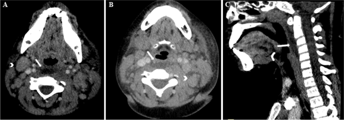
Despite antibiotic treatment, her fever continued, as well as the neck swelling. A cervical CT was performed on the 4th day of fever, demonstrating left retropharyngeal swelling with a hypodense lesion of 45 mm with a suspected abscess (Fig. 1A-C), as well as multiple enlarged and inflamed lymph nodes. Expansion of antibiotic coverage to ampicillin sulbactam also showed no clinical improvement. Subsequently, she underwent surgical drainage, which showed minimal serous fluid and sterile bacterial cultures. A third antibiotic change to Ertapenem, was also without any clinical improvement. On the 8th day of fever, symmetrical arthritis of the PIPs, MCPs, and MTPs joints appeared. On the 9th day of fever, non-purulent conjunctivitis, as well as a strawberry tongue with swollen dry lips were noted. An ophthalmological examination showed mild anterior uveitis. Echocardiography was normal.
At this point, she was diagnosed with Kawasaki disease, and the administration of a single dose of IVIG (2 Gram\kg) led to a rapid clinical improvement and resolution of fever and other signs and symptoms. At her follow-up visit two weeks later, she displayed a desquamation of the skin of the tips of the thumb and third finger. At the follow-up four weeks later, the echocardiography of the coronary arteries was normal.
Discussion
More than 50% of KD patients have atypical presentations that may result in delayed or missed diagnosis and, consequently, high rates of vascular damage. We present in this paper case of KD that presented with a retropharyngeal abscess-like lesion, which is a rare presentation.
Previous literature has shown that 90% of KD patients present with variable head and neck manifestations such as facial exanthema, conjunctivitis, oral mucosal changes, and pharyngitis. Less common presentations are otitis media, torticollis, deep neck infection-like symptoms, meningismus, and acute tonsillitis have been described [7].
Lymphadenopathy as a KD criterion is the least common diagnostic feature (50–75%), as the incidence of the other criteria occurs in 80–90% of the cases. Lymphadenopathy is described as the initial presenting symptom in 12%. Deep neck infection-like presentation is described in only less than 5% of all the patients with head and neck manifestations [2, 9, 13]. Signs and symptoms of retropharyngeal involvement, such as stridor, neck pain, and dysphagia, are less common in KD than in retropharyngeal abscesses [7].
Peritonsillar, retropharyngeal and parapharyngeal swelling are uncommonly reported as initial manifestations of KD. The precise pathophysiology of the association of KD with retroparapharyngeal pathology is unclear, with inflammation and edema hypothesized as the main mechanisms [3, 8].
Tashiro et al. described the typical ultrasound appearance of lymphadenopathy in KD patients as multiple hypoechoic–enlarged nodes forming a palpable mass resembling a cluster of grapes. This is in comparison to the appearance of bacterial lymphadenitis, described as a well-defined mass with a large central hypoechoic area surrounded by satellite normal-sized lymph nodes [18].
Pontell et al. reported the first case of KD mimicking a retropharyngeal abscess in 1994 [9, 10]. Glasier et al. reported that there might be an overlapping CT density number between cellulitis, adenopathy, and abscess, which may lead to false interpretations [19].
Poor response to antibiotic therapy and a negative aspirate culture may be additional diagnostic clues to correctly diagnosing KD [2].
Uveitis can be part of KD during the first week of illness; about three-fourths of children are photophobic, a consequence of anterior uveitis, 64 which peaks between 5 and 8 days of illness and is more common in children over 2 years old [20].
Our literature review found sixteen similar reported cases in the last thirty years. We summarized the clinical presentation, imaging findings, surgical and echocardiographic findings, and antibiotics treatment in Table 1.
The KD patients with abscess-like lesions were predominantly males (82%), with an age range from 10 months to 9 years, with a mean age of 5 years, which is considered higher than the average age of patients with KD. The majority of the cases (94%) initially presented with fever and neck swelling, without additional clinical criteria, leukocytosis. Leukocytosis was in all patients with an average of 18,500 WBC/μL. All the cases were initially considered for possible deep-neck bacterial infection, prompting antibiotic therapy and imaging studies. CT\MRI imaging was performed in all the cases, with findings suspicious for retropharyngeal disease, but in the majority, without enhancement. Indeed, low-density cervical lesions demonstrating minimal to no enhancement should raise the possibility of KD.
Intravenous antibiotics were administered in 16 patients (94%), and surgical drainage was attempted in 7 patients (41%), with two 2 cases (11%) of purulent fluid, only one of them being culture positive for Staphylococcus aureus.
As with our patient, the diagnosis of KD was made in these cases when the fever persisted and other characteristic features developed. The diagnosis was delayed beyond nine febrile days in 5 patients (30%). All the patients had a dramatic and fast clinical improvement with resolution of fever within 48 h after IVIG administration; the majority even showed improvement in the first 24 h. Most of the patients responded to one dose of IVIG; in only two cases, two doses of IVIG were administered due to slow response, and in these two cases, the diagnosis was not delayed and, they were diagnosed on the 7th day of fever.
41% of the patients had cardiac manifestations compared to less than 20% in the general pediatrics pediatric population who are diagnosed with Kawasaki disease.
In summary, we present our patient and review 17 cases reported over 30 years (since 1990), presenting with clinical presentation and radiological findings consistent with the presence of a retropharyngeal abscess. Nearly all patients were treated with antibiotics and surgical exploration without improvement. All the cases responded promptly to IVIG therapy. Cardiac involvement was more common than expected in other cases of KD.
Conclusion
Kawasaki disease requires a high index of suspicion and awareness of unusual presentations. In our case and literature review, Kawasaki disease mimicked a retropharyngeal abscess that was refractory to antibiotics and surgical intervention.
Thus, Kawasaki disease should be kept in mind as one of the differential diagnosis of patients with febrile lymphadenitis and/or retropharyngeal abscess who do not respond to antibiotic treatment in the relevant clinical context. This can prevent delay in diagnosis and detrimental sequelae.
Availability of data and materials
Raw data for this study (i.e., specific data extracted from each publication are available upon request).
Abbreviations
- CAA:
-
Coronary artery aneurysm
- CT:
-
Computerized Tomography
- IVIG:
-
Intravenous Immune Globulin
- KD:
-
Kawasaki disease
- MCP:
-
Metacarpophalangeal
- MTP:
-
Metatarsophalangeal
- PIP:
-
Proximal interphalangeal
References
Isidori C, Sebastiani L, Esposito S. A case of incomplete and atypical kawasaki disease presenting with retropharyngeal involvement. Int J Environ Res Public Health. 2019;16(18):3262.
Choi SH, Kim HJ. A case of Kawasaki disease with coexistence of a parapharyngeal abscess requiring incision and drainage. Korean J Pediatr. 2010;53(9):855–8.
Connell JT, Park JH. Acute peritonsillar swelling: A unique presentation for Kawasaki disease in adolescence. BMJ Case Rep. 2018;2018:1–5.
MacHaira M, Tsolia M, Constantopoulos I, Garoufi A, Kaltsa M, Radiotis A, et al. Incomplete Kawasaki disease with intermittent fever and retropharyngeal inflammation. Pediatric Infectious Disease Journal. 2012;31(4):417–8.
Marchesi A, Rigante D, Cimaz R, Ravelli A, Tarissi de Jacobis I, Rimini A, et al. Revised recommendations of the Italian Society of Pediatrics about the general management of Kawasaki disease. Ital J Pediatr. 2021;47:16 (BioMed Central Ltd).
Conti G, Giannitto N, De Luca FL, Salpietro A, Oreto L, Viola I, Ceravolo A, Nicocia G, Sio A, Romeo M, Ceravolo G, Cuppari C, Calabrò MP, Chimenz R. Kawasaki disease and cardiac involvement: an update on the state of the art. J Biol Regul Homeost Agents. 2020;34(4 Suppl. 2):47.
Kritsaneepaiboon S, Tanaanantarak P, Roymanee S, Lee EY. Atypical presentation of Kawasaki disease in young infants mimicking a retropharyngeal abscess. Emerg Radiol. 2012;19(2):159–63.
Aldemir-Kocabaş B, Kicali MM, Ramoʇlu MG, Tutar E, Fitöz S, Çiftçi E, et al. Recurrent Kawasaki disease in a child with retropharyngeal involvement: A case report and literature review. Medicine (United States). 2014;93(29):e139.
Hung MC, Wu KG, Hwang B, Lee PC, Meng CCL. Kawasaki disease resembling a retropharyngeal abscess - Case report and literature review. Int J Cardiol. 2007;115(2):94–6.
Pontell J, Rosenfeld RM, Kohn B. Kawasaki disease mimicking retropharyngeal abscess. Otolaryngology-Head and Neck Surgery. 1994;110(4):428–30.
McLaughlin RB, Keller JL, Wetmore RF, Tom LWC. Kawasaki disease: A diagnostic dilemma. Am J Otolaryngol Head Neck Med Surg. 1998;19(4):274–7.
Rooks VJ, Burton BS, Catalan JN, Syms MJ. Kawasaki disease presenting as a retropharyngeal phlegmon [1]. Pediatr Radiol. 1999;29(11):875–6.
Homicz MR, Carvalho D, Kearns DB, Edmonds J. An atypical presentation of Kawasaki disease resembling a retropharyngeal abscess. Int J Pediatr Otorhinolaryngol. 2000;54(1):45–9.
Lin PW, Lin HC, Tsai CK. Radiology quiz case 1. Arch Otolaryngol Head Neck Surg. 2010;136(8):1506–9.
Langley EW, Kirse DK, Barnes CE, Covitz W, Shetty AK. Retropharyngeal edema: An unusual manifestation of kawasaki disease. J Emerg Med. 2010;39(2):181–5.
Ganesh R, Srividhya SV, Vasanthi T, Shivbalan S. Kawasaki disease mimicking retropharyngeal abscess. Yonsei Med J. 2010;51(5):784–6.
Singh S, Jindal AK, Pilania RK. Diagnosis of Kawasaki disease. Int J Rheum Dis. 2018;21(1):36–44.
Tashiro N, Matsubara T, Uchida M, Katayama K, Ichiyama T, Furukawa S. Ultrasonographic Evaluation of Cervical Lymph Nodes in Kawasaki Disease. Pediatrics. 2002;109(5):e77–e77.
Glasier CM, Stark JE, Jacobs RF, Mancias P, Leithiser RE, Seibert RW, et al. CT and ultrasound imaging of retropharyngeal abscesses in children. Am J Neuroradiol. 1992;13(4):1191–5.
Smith LB, Newburger JW, Burns JC. Kawasaki syndrome and the eye. Pediatr Infect Dis J. 1989;8(2):116–8.
Acknowledgements
Not applicable.
Conflict of interest
Disclosures (includes financial disclosures): All the authors have no conflicts of interest to disclose.
Funding
No funding was secured for this article to disclose.
Author information
Authors and Affiliations
Contributions
Dr. RKAS, Dr. JMVM, Dr. NN and Dr. MHS coordinated and collected the data, drafted the initial manuscript, reviewed and revised the manuscript. All authors approved the final manuscript as submitted and agree to be accountable for all aspects of the work.
Corresponding author
Ethics declarations
Ethics approval and consent to participate
Not applicable.
Consent for publication
Not applicable.
Competing interests
Not applicable.
Additional information
Publisher’s Note
Springer Nature remains neutral with regard to jurisdictional claims in published maps and institutional affiliations.
Rights and permissions
Open Access This article is licensed under a Creative Commons Attribution 4.0 International License, which permits use, sharing, adaptation, distribution and reproduction in any medium or format, as long as you give appropriate credit to the original author(s) and the source, provide a link to the Creative Commons licence, and indicate if changes were made. The images or other third party material in this article are included in the article's Creative Commons licence, unless indicated otherwise in a credit line to the material. If material is not included in the article's Creative Commons licence and your intended use is not permitted by statutory regulation or exceeds the permitted use, you will need to obtain permission directly from the copyright holder. To view a copy of this licence, visit http://creativecommons.org/licenses/by/4.0/. The Creative Commons Public Domain Dedication waiver (http://creativecommons.org/publicdomain/zero/1.0/) applies to the data made available in this article, unless otherwise stated in a credit line to the data.
About this article
Cite this article
Kasem Ali Sliman, R., van Montfrans, J.M., Nassrallah, N. et al. Retropharyngeal abscess-like as an atypical presentation of Kawasaki disease: a case report and literature review. Pediatr Rheumatol 21, 34 (2023). https://doi.org/10.1186/s12969-023-00812-z
Received:
Accepted:
Published:
DOI: https://doi.org/10.1186/s12969-023-00812-z

